




data visualization + art
Creating the Molecular Case Studies Cover
If your photos aren’t good enough, then you’re not close enough
— Robert Capa
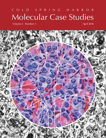
about the cover
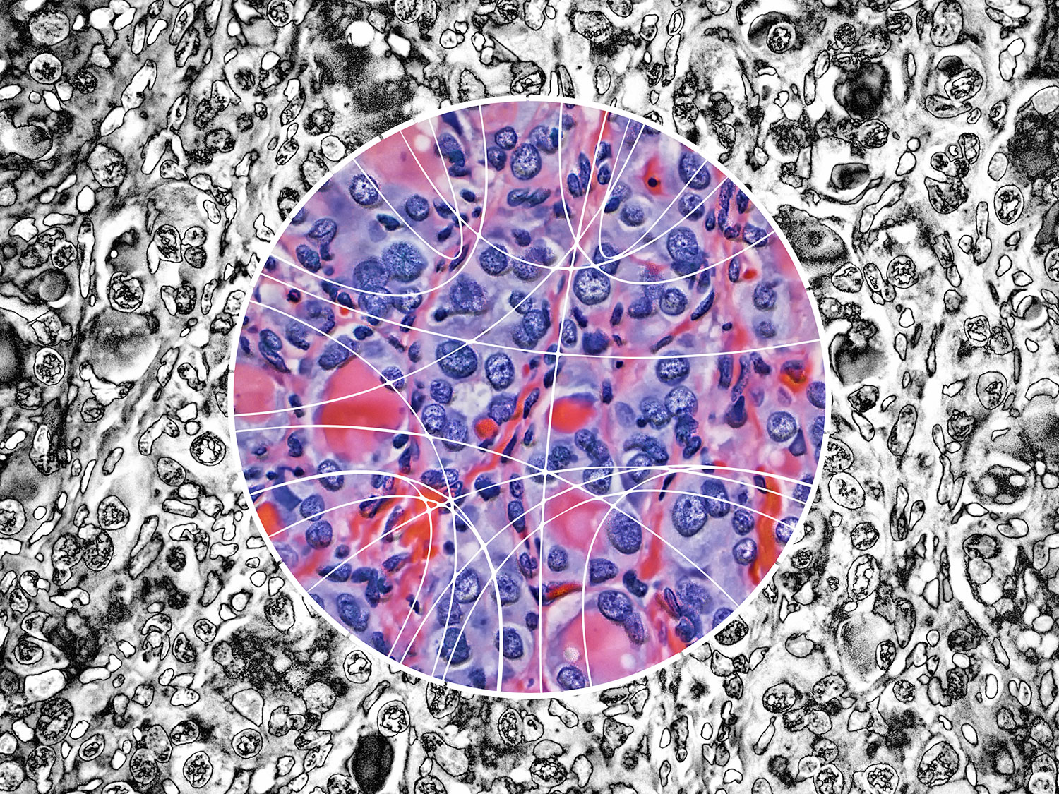
Papillary thyroid carcinoma (PTC) cells, even though malignant, are still genetically programmed to try to be thyroid follicles and may retain their follicular growth pattern, which appear as circles on cross section. Two diagnostic features of papillary thyroid carcinoma are nuclear clearing and intranuclear cytoplasmic inclusions. The black-and-white image is an artistic treatment of a PTC microscopy image (40×) from one of the Personalized Oncogenomics Program study participants at the BC Cancer Research Center. Superimposed is a Circos plot of 17 genomic fusions involving 17 chromosomes identified in the sample by whole-genome sequencing. Showing through the Circos plot is an enhanced color version of the microscopy image. The original image is from Application of genomics to identify therapeutic targets in recurrent pediatric papillary thyroid carcinoma by Ronsley et al. in the April 2018 issue.
The theme of the April issue of Molecular Case Studies is precision oncogenomics. We have three papers in the issue based on work done in our Personalized Oncogenomics Program (POG).
...this special issue provide[s] a glimpse into current cancer precision medicine efforts, reflecting only a microcosm of ... genomics in this bustling space of clinical translation.
John C. Carpten & Elaine R. Mardis
The era of precision oncogenomics
Mol. Case Stud. (2018) 4(2).
I've previously created art based on POG data—posters to celebrate the program's 5-year anniversary.
input materials
The covers of Molecular Case Studies typically show microscopy images, with some shown in a more abstract fashion. There's also the occasional Circos plot.
I've previously taken a more fine-art approach to cover design, such for those of Nature, Genome Research and Trends in Genetics. I've used microscopy images to create a cover for PNAS—the one that made biology look like astrophysics—and thought that this is kind of material I'd start with for the MCS cover.
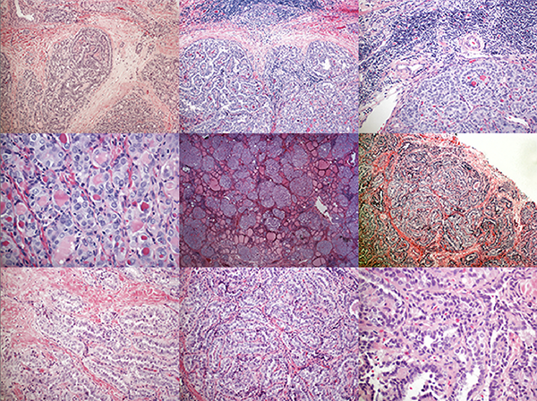
When I look at these kind of images, I have basically no idea what I'm looking at. Sure, I know this is life at tiny scale but I am not a pathologist. This helps me greatly.
Instead, I see color, shapes, and contrast. I hunt for patterns that would make for an interesting visual, without necessarily trying to communicate any of the science behind that—the paper does a much better job at this than I ever could. It's largely a process driven by intuition and my desire to see distinct visual patterns at different length scales with some symmetry, ideally broken in a pleasing way. Vague, I know.
Images of different regions of the same slide, at the same magnification, can have very different levels of visual engagement (for the non-specialist). Just compare the two images below.
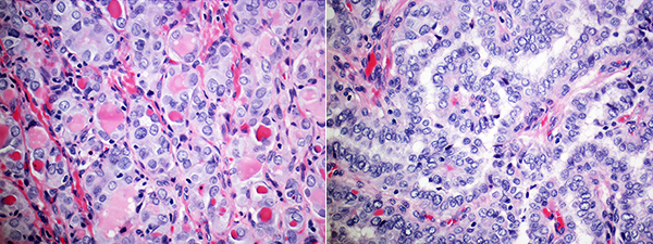
The slide on the left really caught my eye. It had the right proportion of tiny, small, medium and large things.
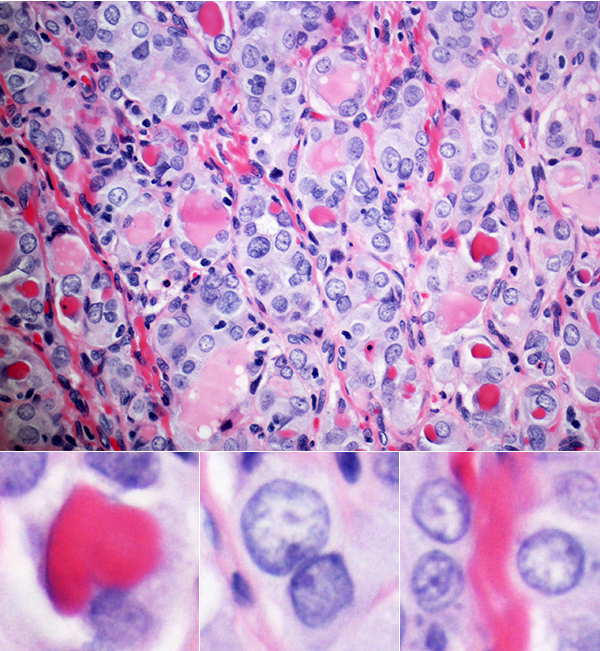
black and white version
The black-and-white version was obtained by solarizing the image. There are both color and black-and-white options for solarization, a method in which various tones of the image are remapped in brightness.
And here's the first black-and-white take.
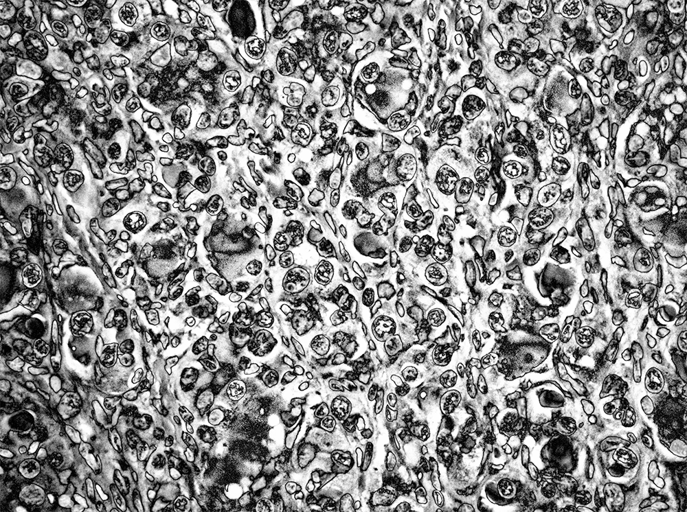
This looked good but a bit dark. I handled this by lightening the tone, differently depending on the element in the image. I also wanted to bring out more details in the internal structure of the cells. This was achieved by applying an otherwise aggressive sharpening mask.
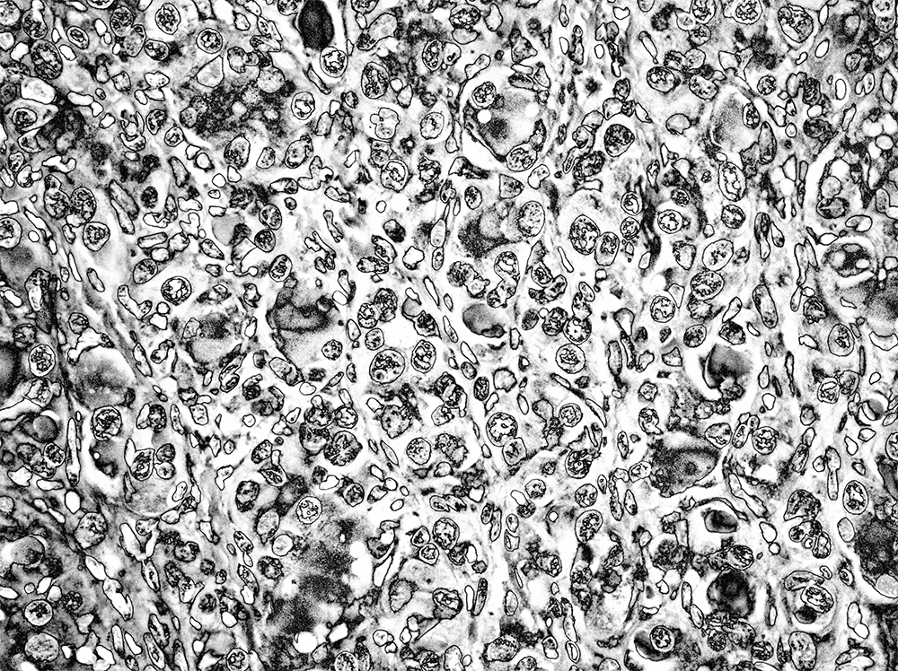
I was quite happy with this result. The combination of solarization and sharpening created a large variety of patterns inside the cells. My brain fought hard to see faces in them.
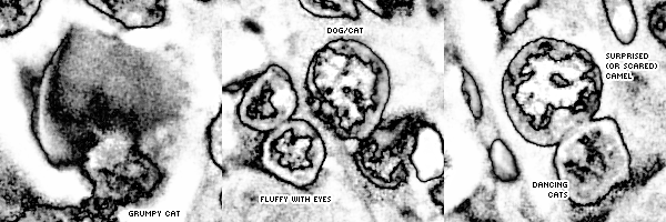
Because I had slides at different magnifications, I created a design in which three slides at 10, 20 and 40 × were composited together so that from left to right the magnification increased across the image. The effect is subtle—you can easily miss it, which is the point.
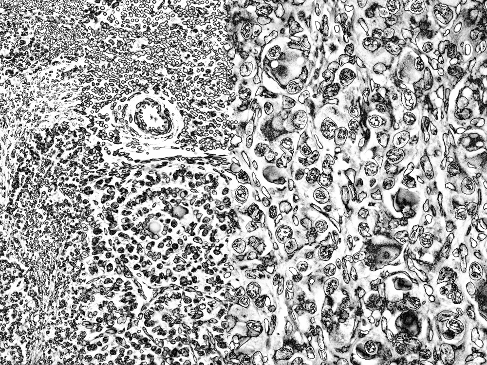
I had pretty high hopes for these black-and-white versions. Previous covers in MCS have been colorful, though, so I thought to provide a color option.
color version
For the color version, I wanted to give the colors more punch. For sure.
I also wanted to emphasize the details, like for the black-and-white image.
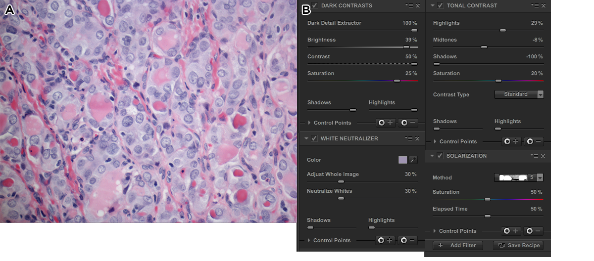
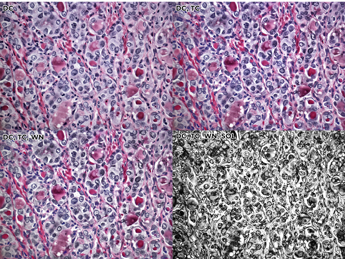
The first process step of the color slide was done using 5 Nik filters, applied in succession: dark contrast, tonal contrast, sunlight, polarization and detail extractor. The effects of the stack of these filters is shown on the original image below. The whole image is shown and in each strip the filters are stacked.
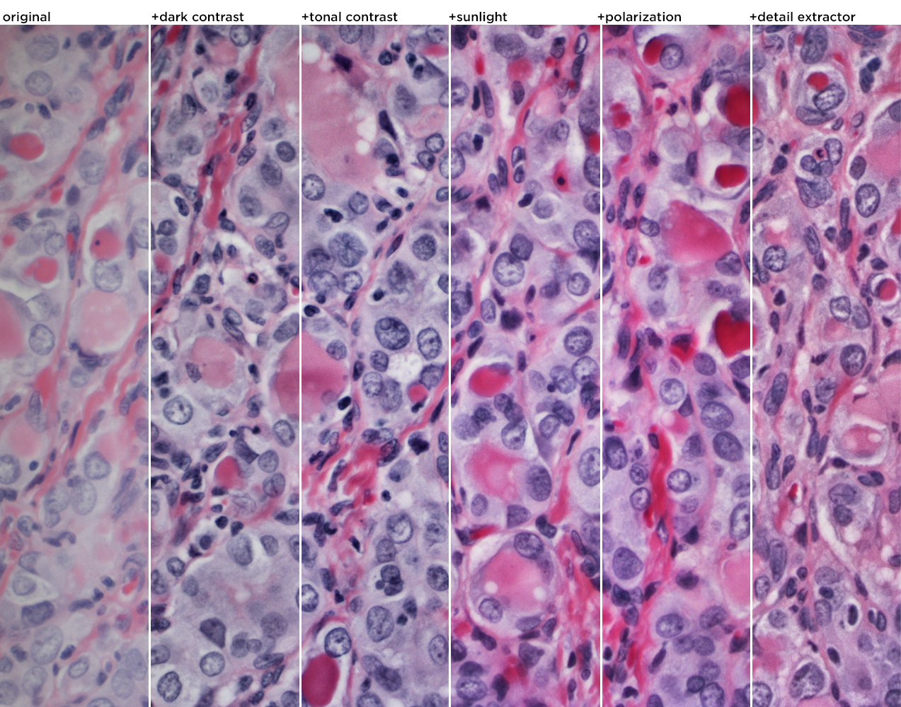
Here's the full image with the 5 Nik filters applied.
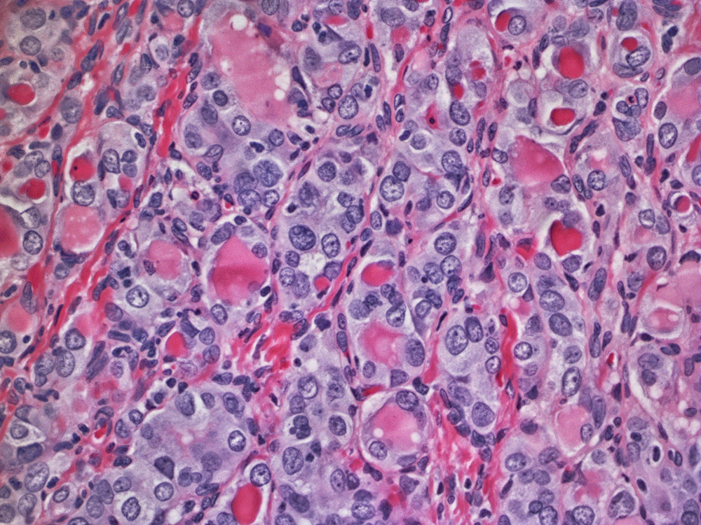
Not there yet, though. I added more sharpening (more than I've ever used before, so I felt a little weird, but got over it quickly). The colors were punched up too—I wanted more contrast between the blue and red areas and transform the reds a little into oranges.
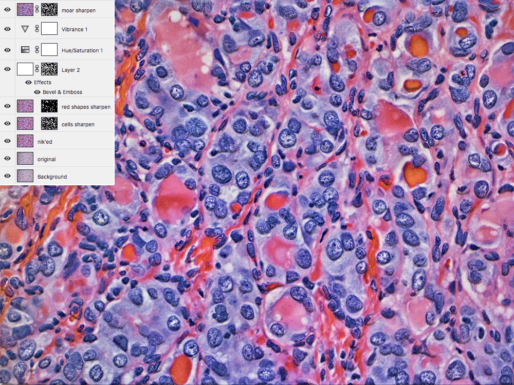
If it looks like the blue areas are popping out of the image, that's the effect of the emboss filter.
final composition
The editors asked me to encorporate a Circos image in the final design. This was tricky—I had spent a lot of time up to now fiddling with extracting patterns and textures from the images.
Something as geometrical and rational as a data graphic would alter the personality of the design. But, the goal of artistic collaboration is always to find a way, so I took some gene fusions that were found in the sample with our structural variant pipeline and created a bare-bones Circos image out of them.
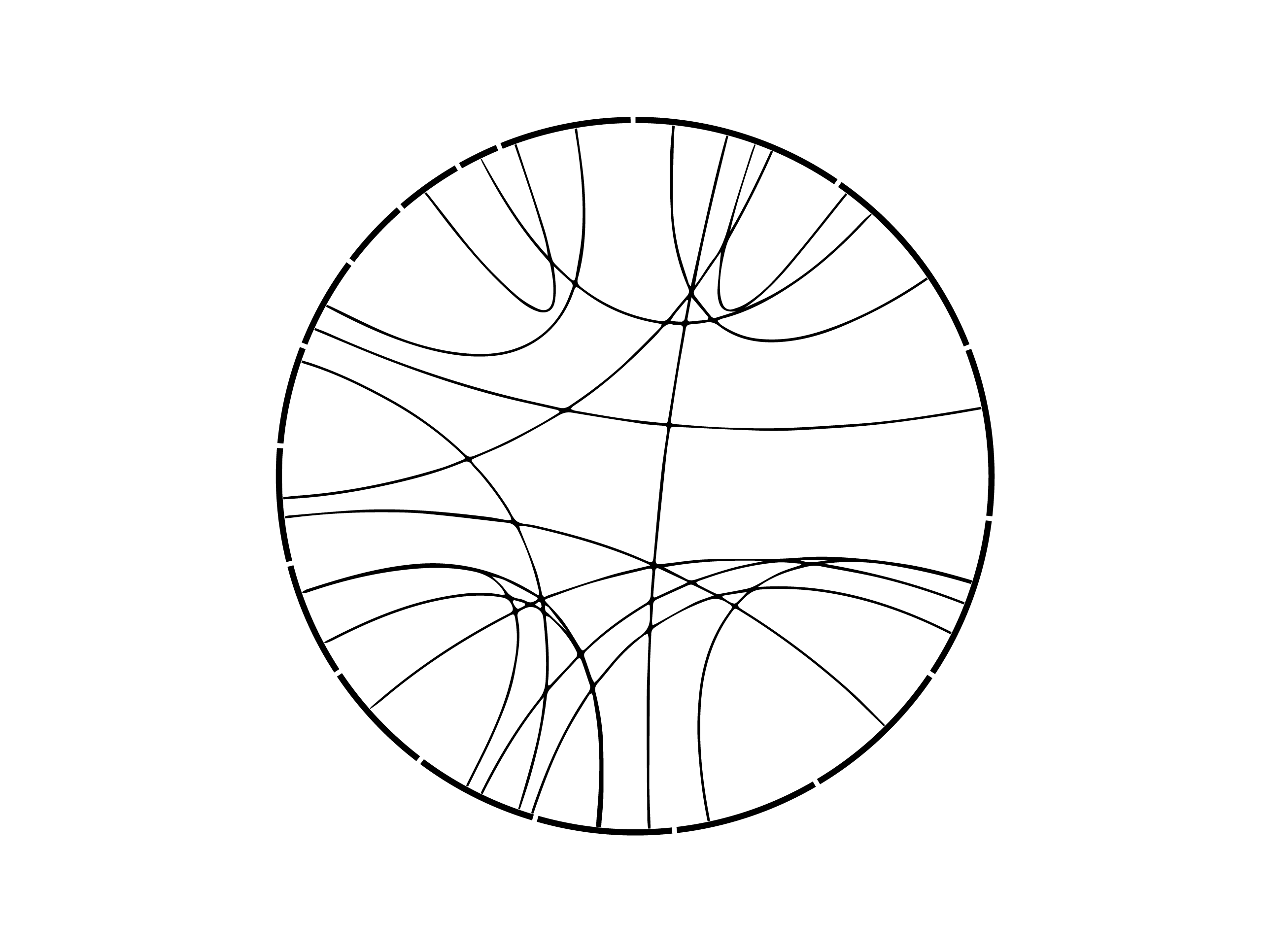
This was then superimposed on the image and emphasized by using the color design inside the circle and black-and-white design outside.
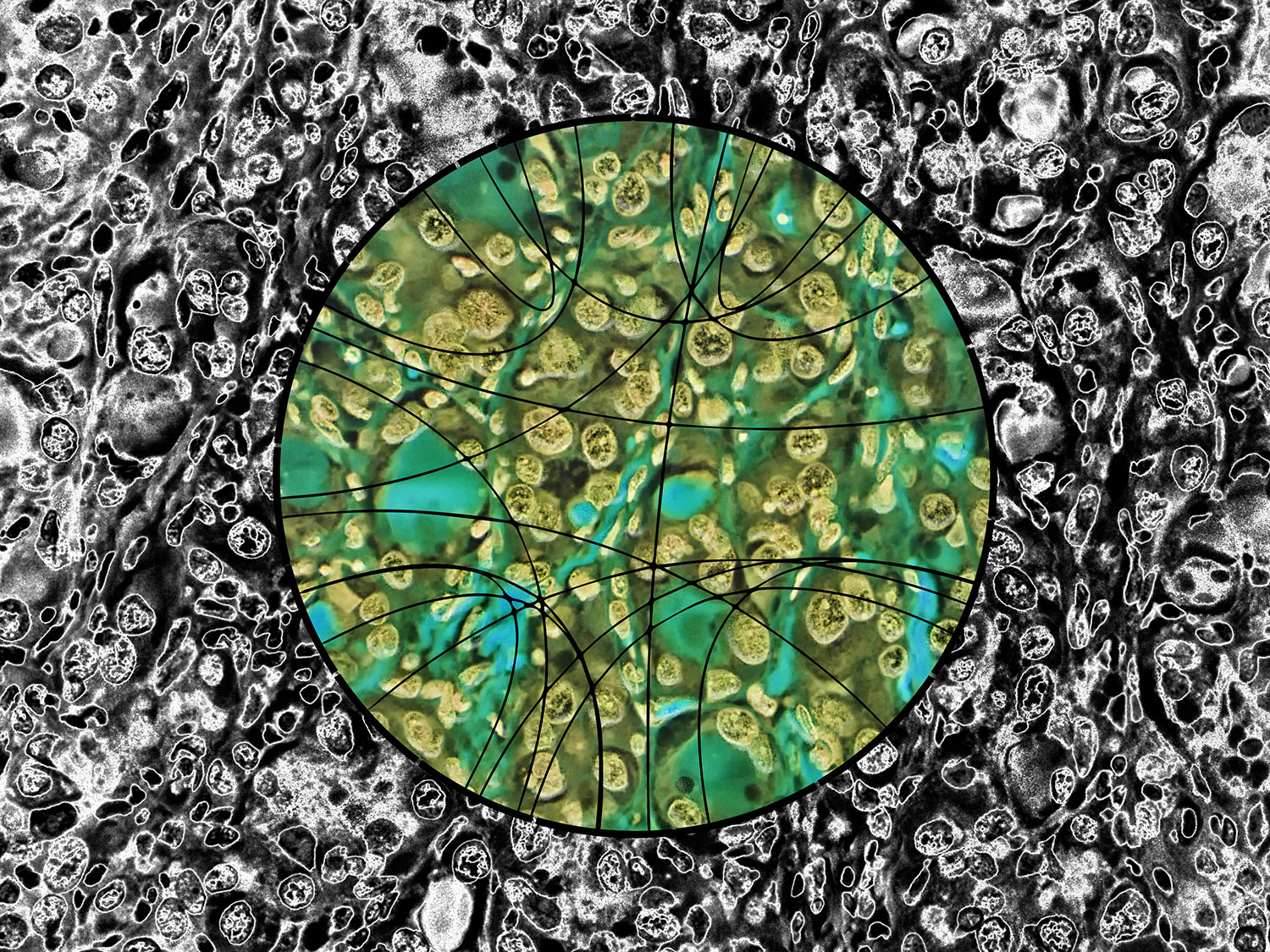
It's always fun to invert images and see what happens.
Beyond Belief Campaign BRCA Art
Fuelled by philanthropy, findings into the workings of BRCA1 and BRCA2 genes have led to groundbreaking research and lifesaving innovations to care for families facing cancer.
This set of 100 one-of-a-kind prints explore the structure of these genes. Each artwork is unique — if you put them all together, you get the full sequence of the BRCA1 and BRCA2 proteins.

Propensity score weighting
The needs of the many outweigh the needs of the few. —Mr. Spock (Star Trek II)
This month, we explore a related and powerful technique to address bias: propensity score weighting (PSW), which applies weights to each subject instead of matching (or discarding) them.
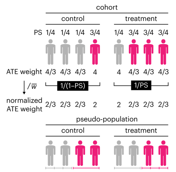
Kurz, C.F., Krzywinski, M. & Altman, N. (2025) Points of significance: Propensity score weighting. Nat. Methods 22:1–3.
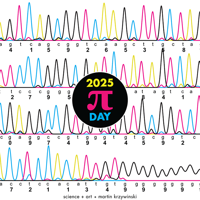
Happy 2025 π Day—
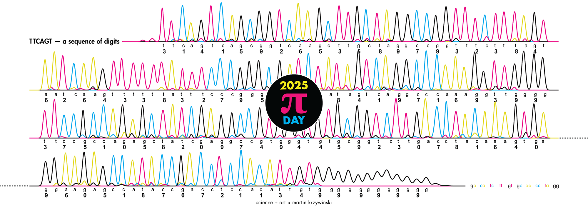
TTCAGT: a sequence of digits
Celebrate π Day (March 14th) and sequence digits like its 1999. Let's call some peaks.
Crafting 10 Years of Statistics Explanations: Points of Significance
I don’t have good luck in the match points. —Rafael Nadal, Spanish tennis player
Points of Significance is an ongoing series of short articles about statistics in Nature Methods that started in 2013. Its aim is to provide clear explanations of essential concepts in statistics for a nonspecialist audience. The articles favor heuristic explanations and make extensive use of simulated examples and graphical explanations, while maintaining mathematical rigor.
Topics range from basic, but often misunderstood, such as uncertainty and P-values, to relatively advanced, but often neglected, such as the error-in-variables problem and the curse of dimensionality. More recent articles have focused on timely topics such as modeling of epidemics, machine learning, and neural networks.
In this article, we discuss the evolution of topics and details behind some of the story arcs, our approach to crafting statistical explanations and narratives, and our use of figures and numerical simulations as props for building understanding.
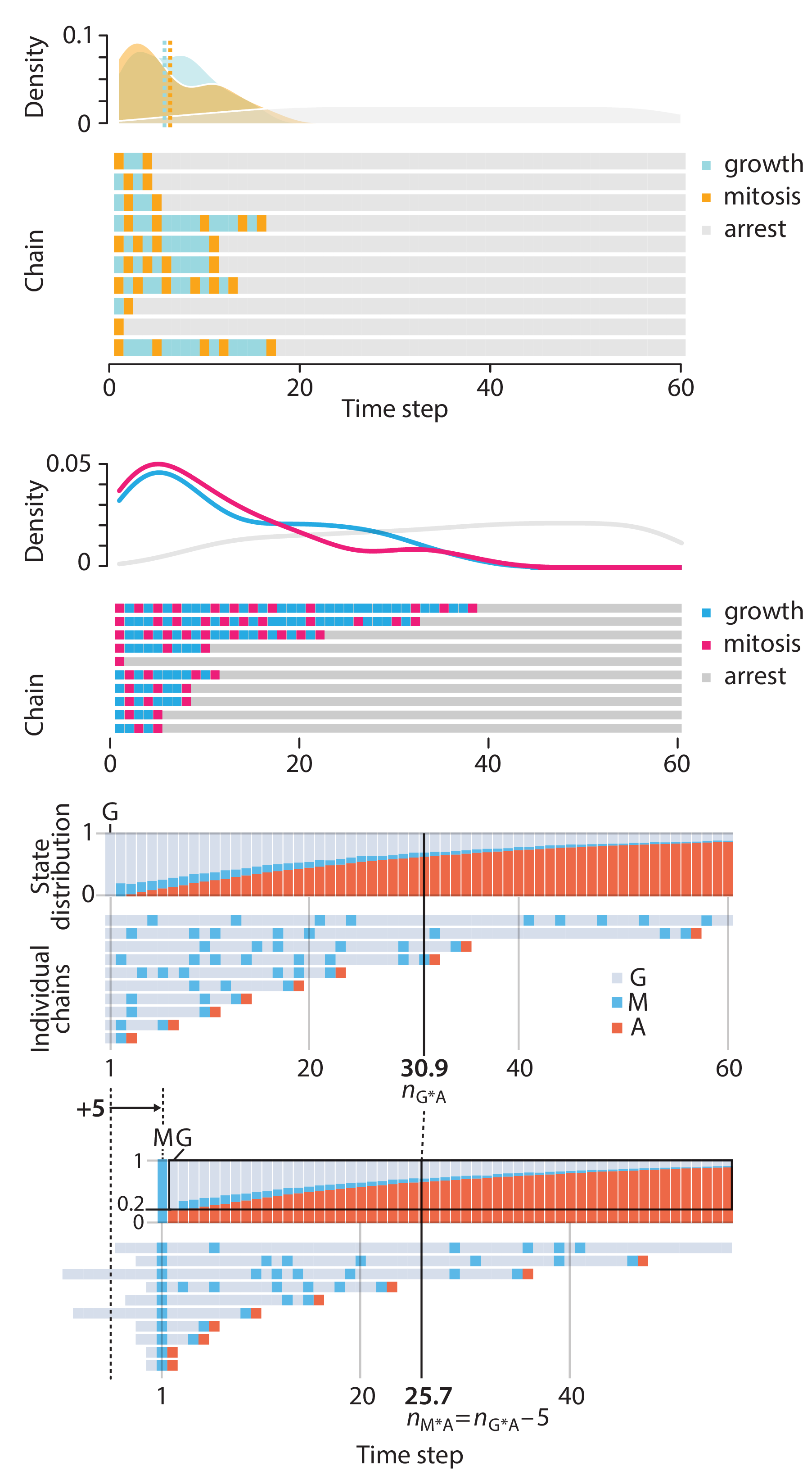
Altman, N. & Krzywinski, M. (2025) Crafting 10 Years of Statistics Explanations: Points of Significance. Annual Review of Statistics and Its Application 12:69–87.
Propensity score matching
I don’t have good luck in the match points. —Rafael Nadal, Spanish tennis player
In many experimental designs, we need to keep in mind the possibility of confounding variables, which may give rise to bias in the estimate of the treatment effect.
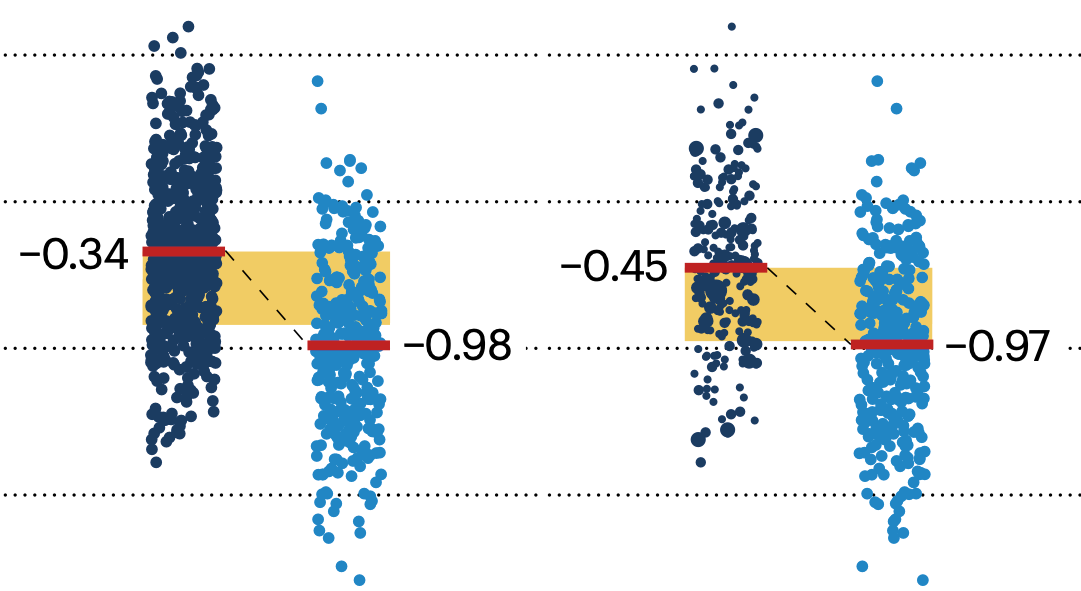
If the control and experimental groups aren't matched (or, roughly, similar enough), this bias can arise.
Sometimes this can be dealt with by randomizing, which on average can balance this effect out. When randomization is not possible, propensity score matching is an excellent strategy to match control and experimental groups.
Kurz, C.F., Krzywinski, M. & Altman, N. (2024) Points of significance: Propensity score matching. Nat. Methods 21:1770–1772.
Understanding p-values and significance
P-values combined with estimates of effect size are used to assess the importance of experimental results. However, their interpretation can be invalidated by selection bias when testing multiple hypotheses, fitting multiple models or even informally selecting results that seem interesting after observing the data.
We offer an introduction to principled uses of p-values (targeted at the non-specialist) and identify questionable practices to be avoided.
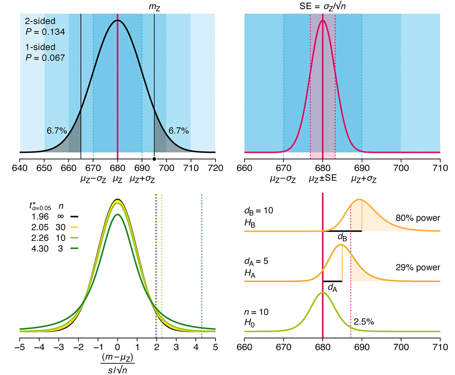
Altman, N. & Krzywinski, M. (2024) Understanding p-values and significance. Laboratory Animals 58:443–446.